Understanding the Technical Differences Between Sonogram and Ultrasound
Many people ask, what is a sonogram and what is an ultrasound? Picture a patient going to a clinic and hearing both words during a checkup. The staff talks about Sonogram vs Ultrasound, but the patient still feels confused. In easy words, ultrasound is the test that uses sound waves. A sonogram is the picture made from this test. When someone asks, “what is a sonogram,” they mean the image. When they ask, “what is an ultrasound,” they mean the scan itself.
Key Takeaways
Ultrasound is a medical test that uses sound waves. It makes pictures of the inside of the body. A sonogram is the picture made by the ultrasound test. It shows organs, tissues, or a baby. Ultrasound is safe and does not hurt. It does not use harmful radiation. This makes it good for people of any age. Real-time ultrasound shows moving pictures. This helps doctors see organs working. It also helps doctors guide some procedures. Sonographers do the ultrasound test and take sonogram pictures. Technicians help with the process. Using clear words helps patients understand their care. Ultrasound means the test. Sonogram means the picture. There are different types of sonograms. These include 2D, 3D, 4D, and Doppler. Each type gives different views and details. This helps doctors diagnose and monitor health. Good communication and records help patient care. They also stop confusion during doctor visits.
Sonogram vs Ultrasound
Key Differences Between Sonogram and Ultrasound
Procedure vs. Image
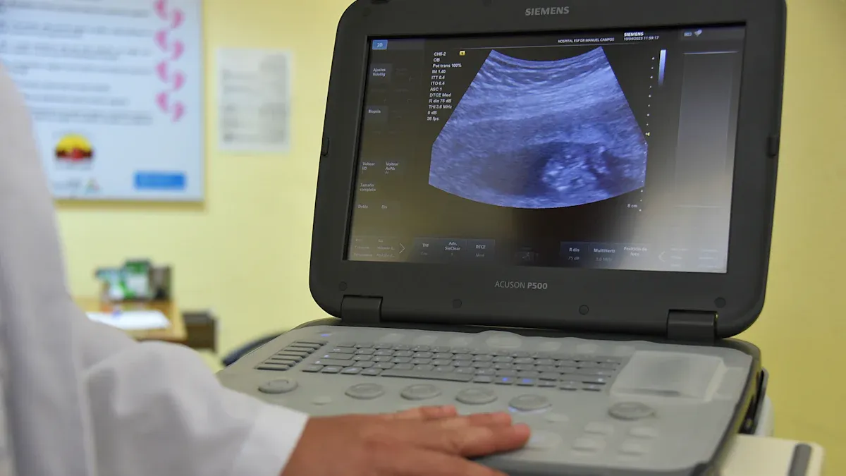
Doctors talk about sonogram and ultrasound, but they are not the same. An ultrasound is a test that uses sound waves to look inside the body. First, the patient gets ready for the test. A special gel is put on the skin. Then, a tool called a transducer moves over the skin. This tool sends sound waves into the body. The sound waves bounce back as echoes. The machine uses these echoes to make pictures.
A sonogram is the picture made from the ultrasound. The sonogram shows what is inside the body. It can show a baby, the liver, or the heart. Healthline and University Health Lakewood Medical Center say ultrasound is the test, and sonogram is the picture. Medical books and rules also explain this difference.
To sum up, ultrasound is the test, and sonogram is the picture.
Terminology in Practice
Doctors, nurses, and sonographers use these words carefully. In medical imaging, ultrasound means the test or technology. Sonogram means the picture or result. For example, a doctor may say, “The ultrasound showed a healthy heart.” This means the test found no problems. The doctor may also say, “The sonogram looks normal.” This means the picture looks fine.
Term | What It Means | Example in Practice |
|---|---|---|
Ultrasound | The imaging procedure using sound waves | "The patient had an ultrasound." |
Sonogram | The image produced by the ultrasound procedure | "The sonogram shows the baby." |
Doctors and nurses keep this difference clear to avoid confusion. They use ultrasound for the test and sonogram for the picture. This is important when talking about results or writing reports.
Are Ultrasound and Sonogram the Same Thing
Common Misconceptions
Many people ask if ultrasound and sonogram are the same. This question comes up a lot in hospitals and clinics. Some patients think these are two different tests. Others think ultrasound uses harmful radiation or is only for pregnancy. These ideas are wrong.
Many people think sonogram and ultrasound are two different things, but ultrasound is the test and sonogram is the picture.
Some believe ultrasound uses radiation, but it does not. Ultrasound uses sound waves, which are safe.
People often think ultrasound is only for babies, but doctors use it for the heart, blood vessels, and muscles too.
Some are confused about how fast results come. Sonogram pictures often show up right away during the scan.
These wrong ideas can make people worry or cause problems when talking to doctors.
Technical Clarification
Medical groups and books agree on the main differences between sonogram and ultrasound. Ultrasound is the test or technology that uses sound waves to make pictures of the inside of the body. Sonogram is the picture made by the ultrasound test. Sonography means using ultrasound for medical pictures.
Knowing the difference between sonogram and ultrasound helps everyone talk clearly. When people use the right words, it is easier to talk about test results and treatment.
Clear words are important in medical imaging. When a doctor says an ultrasound will be done, the patient knows a test will happen. When the doctor talks about the sonogram, the patient knows it is about the picture. This helps stop confusion and makes care better.
How Ultrasound Works

Ultrasound Imaging Basics
Sound Waves and Echoes
Ultrasound imaging works using a simple science idea. The machine sends sound waves into the body. These sound waves come from crystals inside the probe. The waves move through the body and bounce back when they hit something different, like an organ or bone. The probe catches these echoes and turns them into signals. The machine uses these signals to make pictures of the inside of the body.
Ultrasound imaging uses sound waves made by the piezoelectric effect in the probe’s crystals.
These sound waves move through the body and bounce back at places where tissue changes.
The probe picks up the echoes and turns them into signals.
The machine uses these signals to make pictures of what is inside.
This works by sending, bouncing, and catching sound waves as they meet different tissues.
Medical ultrasound uses sound waves that are much higher than people can hear. These waves range from about 1 MHz to 50 MHz. People can only hear up to 20 kHz. The high frequency makes the pictures clearer but does not go as deep. Other scans, like X-rays or MRI, do not use sound waves.
Real-Time Imaging
A big strength of ultrasound is real-time imaging. The machine shows moving pictures as the probe moves on the skin. This helps doctors see organs working, like a heart beating or a baby moving. Real-time pictures also help doctors during some procedures, like taking a tissue sample.
Equipment and Procedure
The Role of the Probe
The probe is the most important part of the ultrasound machine. It sends out sound waves and catches the echoes. The probe uses the piezoelectric effect to change energy into sound waves and back. Different probes help doctors look at different body parts.
A regular ultrasound machine has several main parts:
Transducer Probe: Sends and catches sound waves.
Central Processing Unit (CPU): Makes signals into pictures.
Display Unit: Shows pictures for doctors to see.
Keyboard with Control Knobs: Lets users enter information and change pictures.
Printer: Prints pictures for records.
Safety and Non-Ionizing Radiation
Ultrasound is a safe way to make medical pictures. It does not use ionizing radiation like X-rays or CT scans. The sound waves may cause small heating and movement in tissues, but studies show no serious problems in normal use. Doctors follow safety rules to make sure ultrasound is safe. Groups around the world have rules to keep ultrasound safe for everyone.
Note: Ultrasound is the safest imaging choice for children, pregnant women, and people who need many scans.
Understanding Sonograms
What a Sonogram Shows
A sonogram is a picture of the inside of the body. When people ask, "what is a sonogram," they want to know what the picture shows. Doctors use sonograms to look at organs and tissues. They can also see blood moving in the body. For example, a sonogram can show the liver, kidneys, heart, or a baby growing. Sometimes, doctors use real-time sonograms to help guide needles for anesthesia. This helps them stay away from nerves and blood vessels.
A sonogram can show many normal and abnormal things, like:
Veins and arteries
Nerves and lymph nodes
Doctors use sonograms to find problems such as:
Swelling or infection in tissues
Cysts and abscesses
Tumors or abnormal growths
Blocked blood vessels or blood clots
A sonogram helps doctors find out why someone has pain, swelling, or injury. It can also check for kidney stones, gallstones, or liver problems.
Static vs. Real-Time Images
A sonogram can be a still picture or a moving one. A still sonogram is like a photo. A real-time sonogram shows things moving, like a heart beating or a baby kicking. Real-time pictures help doctors see how organs work and guide them during some procedures.
Tip: Real-time sonograms are great for watching blood flow or helping guide a needle safely.
Interpreting Results
Doctors and sonographers must look at sonograms very carefully. They check for normal shapes and patterns. Sometimes, mistakes happen if the picture is not clear or the person reading it is not trained well. Common mistakes include missing small changes, mixing up normal and abnormal things, or not following the right steps.
To help stop mistakes, medical teams:
Train sonographers often
Use better machines that make clearer pictures
Follow set steps for scanning
Get patients ready, like asking them to fast if needed
Talk well with each other
When someone asks, "what is a sonogram," it is important to know the answer depends on how good the picture is and how well it is read.
Types of Sonograms
2D, 3D, and 4D
Doctors use different sonograms for different reasons. The most common is the 2D sonogram. It shows a flat, black-and-white picture. This type helps check for pregnancy, organ health, and many problems.
Some clinics have 3D sonograms. These make three-dimensional pictures that show more detail. Parents like 3D sonograms because they can see a baby's face or hands. Doctors use 3D sonograms to find birth defects or bone problems.
A 4D sonogram adds movement to the 3D picture. It looks like a video. This lets parents watch a baby move in real time. Doctors sometimes use 4D sonograms to see how organs work, but families mostly use them for keepsake videos.
Ultrasound Type | Image Quality | Diagnostic Utility | Additional Notes |
|---|---|---|---|
2D Ultrasound | Flat, black-and-white, less detail | Main tool for checking health and finding problems | Most common, covered by insurance |
3D Ultrasound | Static, three-dimensional, more detail | Shows faces, bones, and birth defects | Used for bonding and some diagnoses |
4D Ultrasound | Moving 3D images, like a video | Shows movement and organ function | Mostly for keepsakes, some medical uses |
Doppler Sonograms
A Doppler sonogram shows how blood moves in the body. It can measure how fast and which way blood flows. Doctors use Doppler sonograms to find blocked arteries, blood clots, or weak blood flow. This type helps check the heart, veins, and blood supply to a baby during pregnancy.
Doppler sonograms come in different types:
Color Doppler shows blood flow in color.
Power Doppler finds slow flow in small vessels.
Spectral Doppler graphs blood flow speed.
Duplex Doppler mixes pictures and flow data.
A Doppler sonogram gives doctors more details than a regular sonogram. It helps plan treatment and watch heart or blood vessel problems.
Ultrasound vs. Sonogram in Medical Practice
Common Applications
Obstetrics and Prenatal Care
Doctors use ultrasound and sonogram images a lot in pregnancy care. These tools help doctors know if someone is pregnant. They also check how healthy the baby is and how far along the pregnancy is. Doctors look for problems with chromosomes and check the baby's organs. Doppler ultrasound checks how blood moves in the placenta and umbilical cord. Newer 3D and 4D sonograms let parents watch their baby move in real time. These scans give doctors facts about how the baby is growing and feeling.
Cardiology and Vascular Imaging
Doctors use ultrasound to look at the heart and how it works. Echocardiography uses ultrasound to show the heart beating right now. This helps doctors find problems with heart valves and measure the heart’s size. It also helps check for heart disease. Vascular ultrasound checks how blood moves in veins and arteries. It helps doctors find blood clots and blocked blood vessels. These tests are important for people who might have a stroke or need surgery.
Abdominal and Musculoskeletal Uses
Ultrasound and sonogram images help doctors check the belly area. They look at the liver, kidneys, gallbladder, and pancreas. These tests help find gallstones, liver problems, kidney issues, and extra fluid in the belly. In muscle and joint care, ultrasound helps find injuries to muscles, tendons, and joints. Sports doctors use sonograms to guide shots and check for tears or swelling. These tests give fast answers and help doctors plan what to do next.
Note: Doctors use ultrasound or sonogram depending on the medical field. Both help doctors find problems faster and make patients feel better.
Medical Field | Common Applications of Ultrasound and Sonogram |
|---|---|
Obstetrics | Confirm pregnancy, check fetal health, screen for birth defects, monitor growth, use Doppler for blood flow, 3D/4D imaging |
Cardiology | Echocardiography, real-time heart imaging, check for valve disease |
Abdominal | View organs, diagnose gallstones, liver and kidney disease, check for fluid |
Musculoskeletal | Diagnose muscle and joint injuries, guide procedures |
Roles of Sonographers and Technicians
Sonographer Responsibilities
Sonographers have an important job during an ultrasound test. They get the room ready and check the machine. They look at the patient’s history and explain what will happen. Sonographers help the patient get into the right position. They use the transducer to take clear sonogram pictures. They make sure the pictures are good and cover all needed areas. Sonographers look for signs of problems and write a report for the doctor. They do not tell the patient what is wrong but give the doctor important details. Sonographers also help patients feel safe and answer questions.
Get the room and machine ready
Check patient history and explain the test
Help the patient get into position and use the transducer
Take and check sonogram pictures for quality
Write down any problems and make a report
Help patients and answer their questions
Follow safety and work rules
Technician Duties
Ultrasound technicians work with sonographers to help with tests. They set up the equipment and help keep track of patient schedules. Technicians keep records and sometimes help during the test. They help patients get into position or bring supplies. Technicians make sure the machines work well and tell someone if there is a problem. Some technicians clean and take care of the exam room. Both sonographers and technicians need special training and certificates. Groups like the American Registry for Diagnostic Medical Sonography give these certificates. They keep learning new things to stay up to date.
Tip: Sonographers take and check sonogram pictures. Technicians help with equipment, patient care, and support the process.
Why the Distinction Matters
Patient Communication
Avoiding Misunderstandings
Using clear words helps patients feel calm during tests. When doctors say ultrasound for the test and sonogram for the picture, patients know what will happen. Many people feel nervous before an ultrasound because they do not know what to expect. Studies show that when doctors explain things simply, patients feel less scared and listen better. This means the pictures are better and the test does not need to be done again.
When doctors use easy words, patients trust them more and feel safe to ask questions. This trust helps everyone work together for the best results.
If doctors and patients do not understand each other, it can cause worry and mistakes. For example, if a patient thinks a sonogram is a different test, they might worry about missing something important. Using the right words also helps patients know that ultrasound is safe and does not use harmful radiation.
Asking the Right Questions
When patients know the difference between ultrasound and sonogram, they can ask better questions. They might ask, “What will the ultrasound show?” or “Can I see my sonogram image?” This helps them take part in their care. Doctors can help by:
Showing pictures or drawings to explain.
Asking open questions to get patients talking.
Checking that patients understand by having them repeat what they heard.
When patients understand, they follow directions better and feel happy with their care. Working together like this means fewer mistakes and better results.
Medical Records and Reporting
Accurate Terminology
Medical records must keep ultrasound tests and sonogram pictures separate. Doctors write reports about the ultrasound and what they found. They save sonogram pictures in the patient’s file. Hospitals have rules to keep these records safe and neat.
Aspect | Ultrasound Procedures | Sonogram Images |
|---|---|---|
Documentation | Written report with findings and interpretation | Permanently stored and archived in patient record |
Coding | Reported using CPT codes | Not submitted with claims, archived only |
Purpose | Supports billing and medical decisions | Supports clinical, legal, and payer requirements |
Reporting Requirements | Detailed report templates required | Storage governed by regulations |
Using the right words in records helps doctors, nurses, and insurance companies know what was done. It also keeps patients safe by making sure all facts are correct and easy to find.
Impact on Care
When doctors use the right words in reports, patient care gets better. Clear records help the team make good choices and stop mistakes. For example, if a report says “ultrasound” but does not have the sonogram pictures, another doctor may not have all the facts needed. Keeping reports and pictures organized also helps with checks, legal reviews, and insurance.
Good communication and clear records help patients trust their doctors. They also make sure every patient gets the right care at the right time.
Summary: Key Differences Between Sonogram and Ultrasound
Quick Reference Table
Knowing the difference between ultrasound and sonogram helps people talk clearly. This is important for both patients and healthcare workers. The table below shows the main differences:
Aspect | Ultrasound | Sonogram |
|---|---|---|
Definition | Medical test that uses sound waves to make pictures | The picture made by the ultrasound test |
Nature | Safe, painless, does not use radiation, used in many ways | The picture you get from the ultrasound test |
Components | Machine has a computer, screen, printer, and transducer | N/A (just the picture) |
How it works | Transducer sends sound waves, gets echoes, computer makes pictures | Picture shows what the sound waves found inside the body |
Uses | Helps doctors look at organs, check babies, study blood flow, and treat some problems | Doctors use the picture to check organs, watch babies grow, and find health problems |
Safety | Very safe, no radiation, used in many places | N/A |
Examples of application | Used for heart, liver, bladder, spleen, gallbladder, uterus, ovaries, thyroid, pancreas, kidneys, and checking babies | Pictures help find infections, injuries, diseases, and show how babies are growing |
Tip: Ultrasound is the scan or test. Sonogram is the picture made during the scan.
When to Use Each Term
It is important to use the right word in medicine. Ultrasound means the whole test. This includes getting ready, putting on gel, and moving the transducer on the body. Doctors and nurses say ultrasound when they talk about planning a test or telling someone what will happen.
Sonogram means the picture from the ultrasound. After the scan, doctors look at the sonogram to check for health problems. In pregnancy, the ultrasound checks the baby, and the sonogram is the picture parents get. Radiologists and heart doctors use the sonogram to study organs and find problems.
Here are some times to use each word:
A doctor plans an ultrasound to look at the liver.
A nurse says the ultrasound does not use radiation.
After the scan, the doctor checks the sonogram for infection.
Parents get a sonogram picture after a pregnancy ultrasound.
Remember: Ultrasound is the test or scan. Sonogram is the picture or result.
Using the right word helps everyone know what is happening. It also keeps records clear and helps doctors give the best care.
Ultrasound is the test that uses sound waves. A sonogram is the picture made from this test. When doctors use clear words, patients feel calm and trust them more. Doctors sometimes say ultrasound is like a camera, and a sonogram is like a photo. This helps people understand the difference. Using kind words and the ASCKS approach makes patients feel safe and comfortable.
Benefit | Description |
|---|---|
Better Understanding | Patients know what will happen and worry less. |
Improved Care | Using the right words helps keep good records. |
Using the correct word makes visits easier and helps everyone work together for the best care.
FAQ
What is the main difference between an ultrasound and a sonogram?
Ultrasound is a test that uses sound waves to see inside the body. Sonogram is the picture made from this test. Doctors use the ultrasound test to get the sonogram image.
Can a sonogram show movement, or is it just a still picture?
A sonogram can be a still picture or show movement. Some sonograms are like photos. Others show things moving, like a heart beating or a baby kicking.
Is ultrasound safe for everyone?
Doctors say ultrasound is very safe. It does not use radiation. Most people, even pregnant women and kids, can have an ultrasound without worry.
Who performs an ultrasound test?
A trained sonographer or ultrasound technician does the test. They use the machine and take the sonogram pictures. A doctor looks at the images and explains what they mean.
Do all ultrasounds produce a sonogram?
Yes, every ultrasound test makes a sonogram. The machine always creates a picture, even if the doctor does not print it or give it to the patient.
Can ultrasound find problems in organs other than during pregnancy?
Yes. Doctors use ultrasound to check the heart, liver, kidneys, blood vessels, and muscles. It helps find many health problems, not just those about pregnancy.
How should patients prepare for an ultrasound?
How you get ready depends on the type of ultrasound. Some tests need you to fast. Others need you to have a full bladder. The doctor or nurse will tell you what to do before the test.
Tip: Always follow the instructions from your healthcare team for the best results.
Can patients get a copy of their sonogram?
Most clinics can give patients a copy of their sonogram. Patients should ask the staff after the test. Some clinics give printed pictures, while others give digital copies.
See Also
A Clear Guide To What Defines An Active Transducer
Beginner’s Guide To Understanding Oscilloscopes And Multimeters
Comparing SDRAM And Asynchronous DRAM: Key Differences
Exploring The Main Differences Among Common Inverter Chips
Essential Differences Between Run And Start Capacitors Explained